Slipping Rib Syndrome
Slipping rib syndrome is an underrecognized cause of lower rib, chest wall, upper abdominal, flank, or back pain. It may occur when the cartilage that helps support the lower ribs becomes loose or unstable. When the rib moves abnormally, it can irritate an intercostal nerve and cause sharp, catching, burning, or stabbing pain.
Patients may feel a clicking, popping, slipping, or catching sensation. Pain may worsen with twisting, coughing, bending, deep breathing, lifting, reaching, rolling in bed, or certain positions. Some patients can reproduce the pain with specific movements. Others have intermittent symptoms that make the condition difficult to identify.
Why Slipping Rib Syndrome Is Often Confusing
Slipping rib syndrome can mimic several other conditions. Pain may feel like gastrointestinal pain, gallbladder pain, kidney pain, abdominal wall pain, thoracic spine pain, rib fracture, intercostal neuralgia, or chest wall inflammation. Because symptoms can occur near the upper abdomen or chest wall, patients may have extensive workups before rib instability is considered.
Standard imaging may not show the issue because the rib may appear normal when the patient is still. X-rays, CT scans, and MRIs are often static. Slipping rib syndrome may become visible only when the rib moves during a provoking maneuver. This is why dynamic assessment can be important in selected patients.
Slipping Rib Syndrome and Hypermobility
Hypermobility can make rib pain more complex. Patients with Ehlers-Danlos syndrome or hypermobility spectrum disorder may have connective tissue laxity that affects joint and cartilage stability. In some patients, this may contribute to rib subluxation, rib-tip pain, chest wall discomfort, muscle guarding, or intercostal nerve irritation.
When hypermobility is present, treatment should not focus only on the painful rib. The evaluation should consider rib mechanics, breathing patterns, thoracic spine movement, abdominal wall tension, connective tissue laxity, muscle compensation, and chronic pain sensitivity. Patients with hypermobility may also need a more careful and gradual approach to rehabilitation and load management.
How MPM Evaluates Slipping Rib Syndrome
MPM begins with a detailed history and physical examination. The clinician reviews where pain occurs, how it feels, what movements provoke it, whether there is clicking or slipping, what imaging has been done, and whether symptoms overlap with abdominal, thoracic, or nerve-related pain.
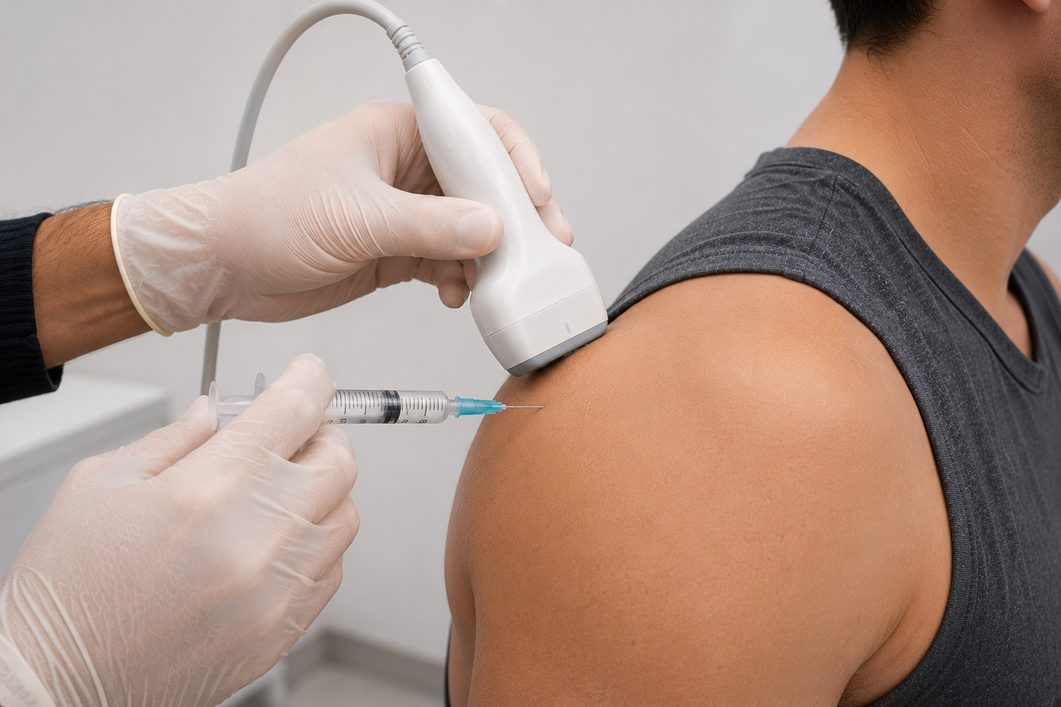
The exam may assess rib tenderness, rib motion, thoracic mechanics, abdominal wall sensitivity, nerve-related symptoms, and pain reproduction. In selected cases, dynamic ultrasound may be used to assess whether rib movement occurs during maneuvers that reproduce the patient’s familiar pain. Diagnostic blocks or ultrasound-guided injections may be considered when the clinical picture supports intercostal nerve irritation or a specific rib-related pain generator.
Dynamic Ultrasound for Slipping Rib Syndrome
Dynamic ultrasound can be useful because slipping rib syndrome is a movement-related condition. The clinician may assess rib motion while the patient performs movements that typically provoke symptoms, such as twisting, bending, coughing, or abdominal contraction. If abnormal rib movement is seen and the patient’s familiar pain is reproduced, this can support the diagnosis.
Dynamic ultrasound is not the only part of the diagnosis. It should be interpreted alongside history, physical examination, red-flag screening, and prior medical evaluation. Rib pain should not be assumed to be slipping rib syndrome until serious cardiac, pulmonary, GI, vascular, infectious, traumatic, or abdominal causes have been considered when appropriate.
Treatment Options for Slipping Rib Syndrome
Treatment depends on severity, anatomy, instability, pain generator, and patient goals. Some patients benefit from activity modification, targeted physical therapy, movement retraining, breathing mechanics, and strategies to reduce provocative rib motion. If intercostal nerve irritation is suspected, ultrasound-guided injections or nerve blocks may be considered in selected cases.
Prolotherapy may be discussed when symptoms suggest ligamentous or cartilage-related instability and the patient is an appropriate candidate. PRP, BMAC, or regenerative medicine may be considered only after careful review of diagnosis, anatomy, risk, and evidence. These treatments should not be presented as guaranteed rib stabilization or cartilage repair.
When Surgery May Be Needed
Some patients with significant rib instability or persistent symptoms may require surgical evaluation. Surgery may be considered when slipping rib syndrome is well supported by the clinical picture and nonsurgical care has not been sufficient. MPM may coordinate referral when symptoms, instability, or functional limitation suggest that surgical assessment is appropriate.
When Rib Pain Needs Urgent Evaluation
Rib, chest, or upper abdominal pain should be evaluated carefully because symptoms can overlap with heart, lung, GI, gallbladder, kidney, vascular, infection, fracture, spine, nerve, and abdominal wall conditions. Patients should seek urgent care for chest pressure, shortness of breath, fainting, fever, coughing blood, severe trauma, severe abdominal pain, unexplained weight loss, new neurologic symptoms, or rapidly worsening pain.
How MPM Approaches Slipping Rib Syndrome Care
MPM approaches slipping rib syndrome through a diagnosis-first, function-focused model. The evaluation considers rib instability, costal cartilage laxity, intercostal nerve irritation, hypermobility, EDS, abdominal wall pain, thoracic spine referral, muscle guarding, and chronic musculoskeletal pain contributors.
For patients looking for slipping rib syndrome treatment in Manhattan, MPM offers a careful pain medicine perspective that can work alongside physical therapy, surgery, orthopedics, and other specialists when needed. The goal is to validate real movement-related rib pain, identify the correct pain generator, and build a treatment plan that fits the patient’s anatomy, instability pattern, risks, and goals.